ARTIFICIAL INTELLIGENCE AND MAMMOGRAPHY
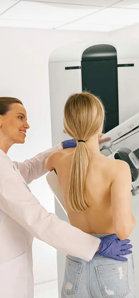
Screening for breast cancer, the most common cancer, is typically done with mammography. Breast MRI and tomosynthesis are also currently used in breast cancer screening. Due to the high-dimensional and complex nature of the resulting images, interpreting them is difficult and time-consuming.
Screening for breast cancer, the most common cancer, is standardly performed with mammography. Breast MRI and tomosynthesis are also used in breast cancer screening today. The resulting images are complex and difficult to interpret, making them time-consuming and difficult. Numerous computer-aided software programs have been developed to increase radiologists' efficiency and accuracy in image analysis for cancer screening.
Due to the success of artificial intelligence systems developed with deep learning methods and convolutional neural networks in image analysis in other fields, artificial intelligence systems have also begun to be used in radiologic image analysis and mammography reading.
One of the benefits of deep learning is the system's ability to learn automatically. Instead of humans teaching image features and the associated calculations to computers, deep learning allows computers to learn image features themselves. In other words, deep learning methods have transitioned from teaching image features to computers learning image features themselves.
The first computer-aided diagnosis (CAD) software was developed in the early 1990s for breast cancer detection in mammography.
CAD systems are crucial for reducing missed or misinterpreted lesions in mammography. Research has shown that using AI-enhanced CAD as a decision support tool helps radiologists more than traditional approaches. Furthermore, research highlights that breast radiologists achieve higher diagnostic performance with AI-enhanced decision support systems compared to reading alone.
It's also important to understand the limitations of AI systems. Machine learning systems, including deep learning systems, can only specialize in solving isolated tasks, while human intelligence makes decisions by synthesizing information from various sources and layers.
AI will certainly impact radiology more rapidly than other medical fields. In just the last few years, various applications have been developed that match or even surpass human performance in certain image recognition tasks, such as breast imaging. The next step is to ensure that these applications become part of daily routines once the necessary approvals and legal frameworks are completed.
In the future, deep learning-based AI systems will increase the efficiency and confidence of breast radiologist's daily routines, freeing up time for radiologists to focus more on the patient's clinical situation. Radiologists will shift their focus from images to the patient with the help of decision support systems developed with AI applications.